Actinic keratosis: Signs and symptoms
What are the signs and symptoms of actinic keratosis?
An actinic keratosis (AK) develops when skin has been badly damaged by ultraviolet (UV) light from the sun or indoor tanning.
Signs of actinic keratosis
The brown spots on this man’s face may look like age spots, but they’re actually actinic keratoses.

Left untreated, some actinic keratoses (AKs) turn into a type of skin cancer called squamous cell carcinoma. That’s why it’s important to know if you have any of these precancerous growths on your skin.
The following pictures show some diverse ways that it can appear.
9 ways a precancerous growth can appear on your skin or lips
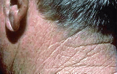
A rough-feeling patch on skin that's had lots of sun. You can often feel an AK before you see it.
A rough-feeling patch on skin
This man said that a small patch of skin on the back of his neck felt like sandpaper. Later, a visible AK appeared on his sandpaper-like patch of skin.
One or more rough, scaly bumps that may look like pimples or spots of irritated skin.
Rough, scaly bumps that may looks like pimples
The arrows on this woman’s face point to actinic keratoses.

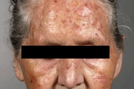
Many scaly, raised spots on the skin that may look like a rash or acne breakout.
Several scaly, raised spots that may look like a rash or acne breakout
Many of the spots on this woman’s forehead, nose, and cheeks are actinic keratoses.
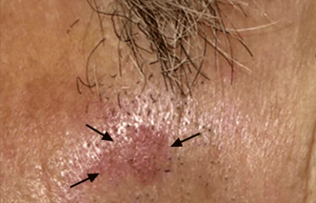
A raised, rough-feeling patch on your skin that may be red, pink, skin-colored, or gray.
A raised, rough-feeling patch that may be red, pink, skin-colored, or gray
The reddish, pink patch below this man’s sideburn is an actinic keratosis.
Flat, scaly area that looks like an age spot. AKs more commonly look like age spots in people who have skin of color.
Flat, scaly area that may look like an age spot
The spot on this man’s nose may look like an age spot, but it’s actually an actinic keratosis. AKs more commonly look like age spots in people who have skin of color.

A dry, scaly lip that never heals (or heals and returns).
A dry, scaly lip that never heals
It may look like this man has a badly chapped lower lip, but the white, dry, and cracked skin is actually a precancerous growth.
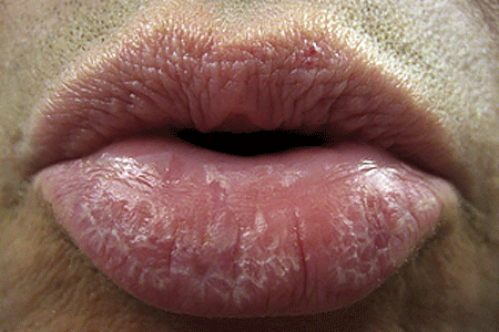
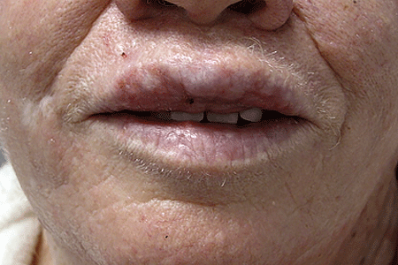
Scaly, white patches on one (or both) lips. White patches on your lips can be a sign that you have a precancerous condition on your lips.
Scaly, white patches on lips
White patches on your lips can be a sign that you have a precancerous condition on your lips.
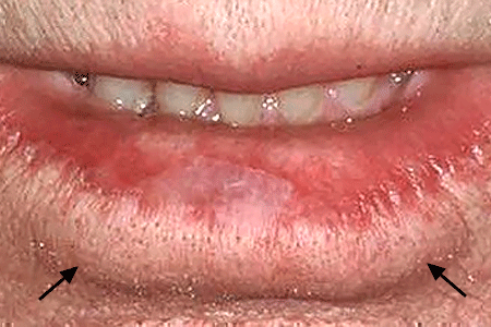
Loss of color on one or both lips.
Loss of color on lips
About half the color on this man’s lower lip has disappeared, turning his lip the same color as the skin on his face.
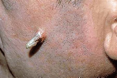
Growth that looks like an animal’s horn. When an AK appears in this form, it often indicates that the person has a higher risk of developing a type of skin cancer called squamous cell carcinoma. A board-certified dermatologist should examine any horn-like growth on your skin ASAP.
A growth that looks like an animal's horn
When an actinic keratosis appears in this form, it often indicates that the person has a higher risk of developing a type of skin cancer called squamous cell carcinoma.
What color is an actinic keratosis?
As the above pictures show, AKs come in many colors. You may see a scaly pimple-like bump or patch of skin that is:
Red or pink
Skin-colored
Gray
Yellow
Brown or tan
White
Does an actinic keratosis hurt?
While most people see only a change to their skin, an AK can:
Itch
Burn or sting
Feel tender or painful when touched
Stick to your clothing, causing discomfort
Bleed
If you find a change on your skin that could be an actinic keratosis, protect your health by seeing a board-certified dermatologist. Should that change be an AK, you have a greater risk of developing skin cancer. Being under the care of a board-certified dermatologist helps to find skin cancer early when its highly treatable.
While having skin that’s been badly damaged by the sun or indoor tanning greatly increases your risk of developing AKs, other things can increase your risk. You’ll find out what else may increase your risk of developing AKs at, Actinic keratosis: Causes.
Related AAD resources
Images
Images 3,4,5,6,7,8,10: Used with permission of the Journal of the American Academy of Dermatology:
Image 3: J Am Acad Dermatol. 2017;76(2):349-50.
Image 4: J Am Acad Dermatol. 2007;56(1):125-43.
Image 5: J Am Acad Dermatol. 2013;69(1):e5-e6.
Image 6: J Am Acad Dermatol. 2010;62(1):85-95.
Images 7, 8: J Am Acad Dermatol. 2012;66(2):173-84.
Image 10: J Am Acad Dermatol. 2000;42(1) part 2:S8-S10.
References
Rigel DS, Cockerell CJ, et al. “Actinic keratosis, basal cell carcinoma, and squamous cell carcinoma.” In: Bolognia JL, et al. Dermatology. (second edition). Mosby Elsevier, Spain, 2008:1645-58.
Vis K, Waalboer-Spuij R, et al. “Validity and Reliability of the Dutch Adaptation of the Actinic Keratosis Quality of Life Questionnaire (AKQoL).” Dermatology. 2018;234(1-2):60-5.
 Atopic dermatitis: More FDA-approved treatments
 Biosimilars: 14 FAQs
 How to trim your nails
 Relieve uncontrollably itchy skin
 Fade dark spots
 Untreatable razor bumps or acne?
 Tattoo removal
 Scar treatment
 Free materials to help raise skin cancer awareness
 Dermatologist-approved lesson plans, activities you can use
 Find a Dermatologist
 What is a dermatologist?


